

New machine learning method: Precise MRI videos despite less measurement data
Scientists at MRI-Lab Graz have produced more precise MRI videos with the help of a new machine learning method and despite blurred training images.
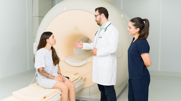
A patient discusses an MRI examination with her doctor.
(Image: antoniodiaz / Shutterstock.com)
Researchers at Graz University of Technology have succeeded in improving the video quality of real-time MRI. Despite fewer and blurred training images, a proprietary machine learning model manages to deliver good images after several passes by adding omitted content. The process can also be mapped to other MRI applications and should save time and money in the future.
A real-time MRI provides a view of the inside when it comes to examining movement sequences. This becomes relevant, for example, when the beating heart or joint and swallowing movements need to be assessed. Compared to static MRI images, doctors can see the situation in real time. However, this is at the expense of image quality.
What the challenge was
The team led by Martin Uecker and Moritz Blumenthal from the Institute of Biomedical Imaging has now succeeded in developing an AI-supported procedure that improves image quality and saves costs and time in the future.
The challenge here is that there are no perfect images as source material for training the neural networks, as are usually used to train AI models. In the field of real-time MRI images, researchers have to rely on a small number of images that are not entirely sharp. To solve this problem, the scientists use the "self-supervised learning" method for their machine learning model. To do this, the team removes a subset of the existing images and has the model add to them until the image matches the removed motifs. The result is more precise real-time images.
Moritz Blumenthal explains the process as follows: "We split the measurement data supplied by the MRI device into two portions. Our machine learning model reconstructs the image from the first, larger data portion. It then attempts to calculate the second portion of the measurement data, which is withheld from it, on the basis of the image." These steps are repeated until the system has learned how to calculate a good image over several attempts.
Not yet in practical use
The application is not yet being used in practice, but can later be used in the field of quantitative MRI, for example for tissue assessment, saving time and money. "This will allow radiologists to access exact data for diagnoses instead of having to interpret images based on differences in brightness based on empirical values," explains Martin Uecker, emphasizing that the new model will also make it possible to speed up long measurements without compromising on quality in the future.
Videos by heise
Felix Nensa, Professor of Radiology with a focus on AI at Essen University Hospital, explains in an interview how artificial intelligence is already being used in radiology. The use of artificially intelligent systems is particularly useful in the field of radiology, for example when it comes to comparing tumor images. Such applications have already been in use for some time as second opinions and indicators.

(hoh)
